Cell- Basic unit of life

The cell is the basic unit of life (smallest unit of a living being).
- It is the structural unit of life because several cells make the structure of an entire organism (e.g. animal body is made of different cells; brain-brain/nerve cells, muscle- muscle cells)
- It is also the functional unit of life because several cells are responsible for proper functioning of an organism as a whole.
Contributors to the study of cell
The cell is the basic structural and functional unit of life.
- Anton Von Leeuwenhoek first saw and described a live cell.
- Robert Hooke first coined the term Cell after observing the compartments in the thin-sliced cork.
- Matthias Schleiden, a German botanist reported that all plants are composed of different kinds of cells which form the tissues of the plant.
- Theodore Schwann (1839), a British Zoologist proposed that; Cells have a thin layer (plasma membrane), Cell wall is unique to the plant cells and the bodies of animals and plants are composed of cells and products of cells.
- Schleiden and Schwann together formulated the cell theory but failed to explain as to how new cells were formed.
- Rudolf Virchow (1855) first explained that cells divide and new cells are formed from pre-existing cells (Omnis cellula-e cellula).
Cell theory
Cell theory was proposed by Schleiden and Schwann. The theory was modified after the introduction of Virchows Omnis cellula-e cellulahypothesis. The unified cell theory states that;
- Cells are building blocks of living organisms i.e. all living organisms are made of one or more cells (Schleiden and Schwann 1838-39)
- All living organisms are composed of cells and cell is the basic structural and functional unit. (Schleiden and Schwann 1838-39)
- All cells arise from pre-existing cells (Rudolf Virchow 1858) i.e. no cell can originate de novo (spontaneously out of nothing)
Significance of cell theory
The cell theory proposed by Schleiden and Schwann is important because
- It provides guidelines (rules) to define life i.e. an organism is believed to have life only if they follow the the three concepts of cell theory
- The theory gave a fundamental (basic) generalization of biology that cell is the smallest unit of all the living organisms (whether unicellular or multicellular).
- The theory also proposed the origin of new cells (cells come pre-existing cells)
Exceptions of cell theory
There are certain organisms and cells which do not agree with the cell theory put forward by Schleiden and Schwann, these are considered to be exceptions and include;
- Viruses are obligate parasites which do not have a cellular machinery but live and multiply in other cells. (No cell structure, but has life)
- Viroids (virus without protein coat), prions (infections protein particles) also behave like viruses and are exceptions to the cell theory
- RBCs and sieve tube cells lack nuclei so, cannot divide and form new cells (new cells are not formed from pre-existing cells)
- Some protozoans and thallophytes (e.g. Acetabularia) have a body but are said to be acellular (body is not made of multiple cells)
- In some organisms, the body is not differentiated into cells though it may have numerous nuclei (coenocytes, e.g., Rhizopus)
Size of the cell

Most of the cells are quite small and can be seen only under a microscope. The human eye cannot see anything smaller than 0.04 mm
- Mycoplasmas, the smallest cells, are only 0.15- 0.3 micrometer in length. Bacteria are around 3- 5 micrometer and human RBC is approximately 7 micrometer. [1 micrometer = 0.001 millimeter (mm)]
- Egg of an ostrich - largest isolated single cell
- Nerve cells are the longest cells.
- Ovum or egg cell is the biggest cell in a womans body.
Bacterial staining
- Bacteria are minute organism with different size and shape.
- Bacterial stain helps in differentiating bacterial species.
- Bacterial smear on the slide is heated slightly and added with crystal violet for a minute.
- Wash and air dry the slide and observe under microscope.
Prokaryotic cell
Prokaryotic cells are the primitive and oldest (present since 3.8 billion years) cells.
They are very small (2-10 microns in size) (1micron= 0.001 millimeters)
They do not have a nucleus.
They have naked DNA (DNA is not covered by nuclear membrane)
They do not have membrane-bound cell organelles.
They have cell wall and ribosomes
Examples include all bacteria (E.coli, Cyanobacteria etc).
Prokaryotic cell

The prokaryotic cells are the cells which do not have a nucleus.
- The DNA/genetic material will be freely floating in the cell cytoplasm.
- These are primitive cells which lack most of the cell organelles (e.g. mitochondria, Endoplasmic reticulum etc.)
- The prokaryotic cells consist of bacteria, blue-green algae, mycoplasma and PPLO (Pleuro Pneumonia Like Organisms).
Shape of the bacteria
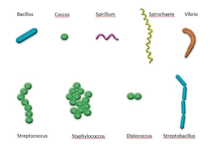
The cell wall of the bacteria gives them different shapes. The bacteria are classified based on their shape as
- Bacilli- Rod-shaped (eg. Bacillus, Lactobacillus)
- Cocci- Spherical shaped (e.g. Staphylococcus, Streptococcus)
- Vibrio- Comma shaped (e.g. Vibrio cholera)
- Spirilla- Spiral/helical shaped (e.g. Spirillum and Helicobacter)
- Diplococci- Cocci arranged in pairs (e.g. Neisseria gonorrhoeae)
- Streptococci- Cocci arranged in in chains (e.g. Enterococcus faecalis)
- Staphylococci Cocci- arranged in bunches (like a bunch of grapes) (e.g. Staphylococcus aureus)
- Tetrads and Sarcinae- Cocci arranged in packs of 4 and 8 respectively. (e.g. Tetrad- Micrococcus, Sarcinae- Sarcina lutea)
- Diplobacilli- Bacilli arranged in pairs (e.g.Coxiella burnetii)
- Streptobacillus- Bacilli arranged in chains (e.g. Streptobacillus moniliformis).
Overview of the structure of a bacterial cell
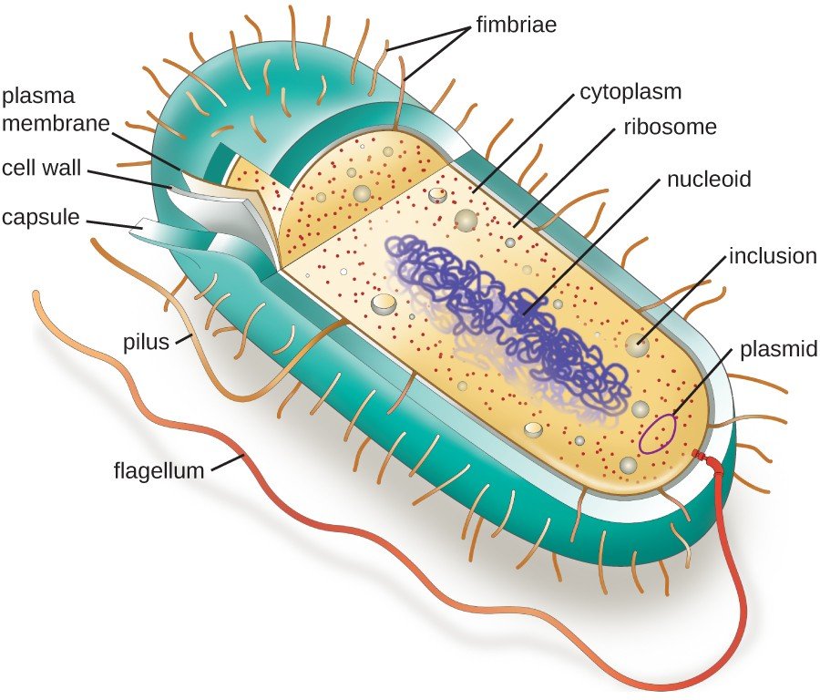
The bacteria are single-celled microorganisms. The structure is made of the following components
- Cell envelope- covers the cell surface
- Mesosome- special membrane structure present in some bacteria
- Cytoplasm- Fluid that fills a cell.
- Nucleoid- naked (not covered) DNA of the cell
- Ribosomes- protein synthesizing components
- Cytoskeleton- responsible for support of the cell.
- Flagella and Pili- Extracellular appendages (attachments) in some bacteria
- Inclusion bodies - stored food (granules)
Ultra structure of the bacterial cell-Cytoskeleton
Cytoskeleton, as the name suggests is the skeleton-like structure of the cell that supports the cell structure.
- It is distributed all over the cytoplasm
- Made of a network of filamentous proteinaceous (made of protein) structures.
- Provides mechanical support
- Important for maintaining the cell shape
- Helps in the motility of the cell
Ultra structure of the bacterial cell- Cell envelope

Cell envelope is the outer most covering of bacteria and consists of 3 layers
Glycocalyx/capsule/sheath/lipopolysaccharide layer
Glycocalyx/capsule/sheath/lipopolysaccharide layer
- Outermost layer in some bacteria.
- It is present as a loose sheath called the slime layer or it is thick and tough, called the capsule.
- Determines the shape of the cell
- Provides a strong structural support
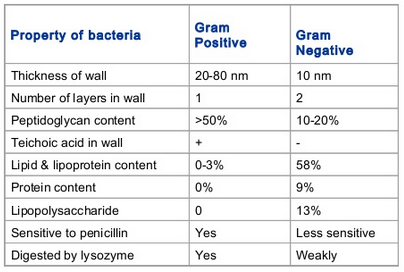
- Made of peptidoglycan, polysaccharide, lipid and protein molecules
- Some bacteria contain Teichoic acid in their cell wall.
- The bacteria are classified into Gram negative and Gram positive based on the structure and composition of the cell wall.
- It serves as a molecular barrier preventing entry of unwanted molecules
- Structurally similar to the Eukaryotic cell membrane.
- The space between the cell wall and plasma membrane is called the periplasmic space
- Made of lipoproteins
- Some bacteria in their PM contain a specialized structure called the Mesosome.
Ultra structure of the bacterial cell- Cytoplasm
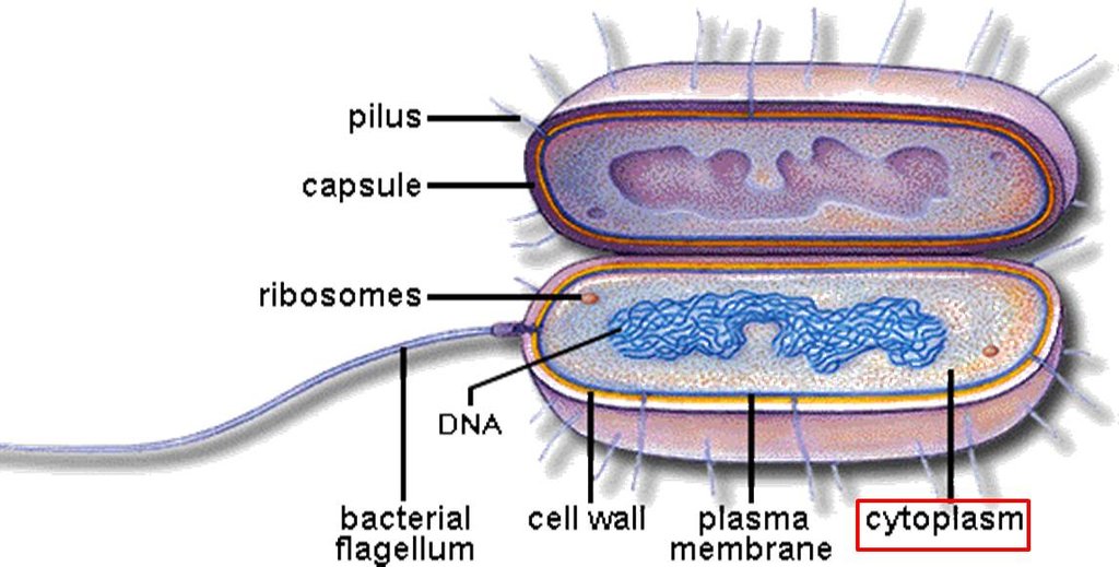
Cytoplasm is also called the cytosol
- It is the fluid matrix that fills the entire cell.
- The Plasmamembrane forms the boundary for the cytosol.It contains all the enzymes required for metabolism.
- All the metabolic reactions of the cell occur in the cytoplasm
Ultra structure of the bacterial cell- Nucleoid

Nucleoid is also called as the Incipient nucleus which is the naked DNA (not covered with a nuclear membrane) in prokaryotes.
- The prokaryotes lack a nucleus.
- They have one double-stranded circular DNA which is also called as genophore.
- It is condensed in the central region of the cell (floating in the cytosol)
- The DNA has close to 2000-3000 genes coding for different proteins
- It is seen attached to the plasma membrane before cell division (attachment is believed to help in separation of the 2 copies of DNA)
Ultra structure of the bacterial cell- Mesosome
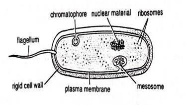
Mesosome is a specialized folding of the cell membrane found in certain bacteria
- The folding of the mesosome consists of vesicles, tubules and lamellae.
- It is Involved in cell wall synthesis, replication of DNA and its distribution to the daughter cells
- It also plays an important role in respiration and secretion process
- By forming folds it increases the surface area of the plasma membrane.
- It is a characteristic feature of prokaryotes (not found in any Eukaryotes)
- In Cyanobacteria, there are membranous extensions into the cytoplasm called Chromatophores which contain pigments that help in photosythesis
Ultra structure of the bacterial cell- Flagella, Pili and Fimbriare

Some bacteria contain extracellular appendages (attachments) which help the bacteria in locomotion, attaching to a surface etc.,
- Motile bacteria contain extensions called flagella, which help in the locomotion process
- A flagellum is a whip-like structure and their number and location on the bacterial surface vary.
- Pili also called sex pili are other extensions with no role in motility.
- A pilus is a hairlike appendage found on the surface of many bacteria. It serves in DNA exchange.
- Pili are made of special protein which also help in the attachment of the bacteria to specific surfaces or to other cells.
- Fimbriae are small bristle-like fibres sprouting from the cell surface in large numbers.
Why are prokaryotic ribosomes 70S not 80S

The ribosomes in the prokaryotes are 70S type (i.e. they have a sedimentation coefficient of 70S) [S= Svedberg unit, which is a unit of sedimentation coefficient. (1S = 1 X 10 sec)]
- The larger subunit of the ribosome is 50s
- The smaller subunit is 30s
- So, together they are supposed to be 80s. But, the prokaryotic ribosomes are 70s because the sedimentation coefficient (S) depends on several factors like; the size, weight and shape of the subunits etc.
- When the (S) value of the smaller and larger subunit are measured they are 50S and 30S (imagine a ball cut into two halves, now it is difficult to roll the two halves of the ball)
- When they combine together their shape changes (now it is round/spherical and can roll easily on the surface).
- The S value is directly proportional to the time taken to move on the surface. So the more time it takes to move the more will be the (S) value
- When the two subunits are together they get a spherical shape and move quickly (takes less time to move) so S value will be lower.
- So, the ribosomes are 70S not 80S
Ultra structure of the bacterial cell- Ribosomes
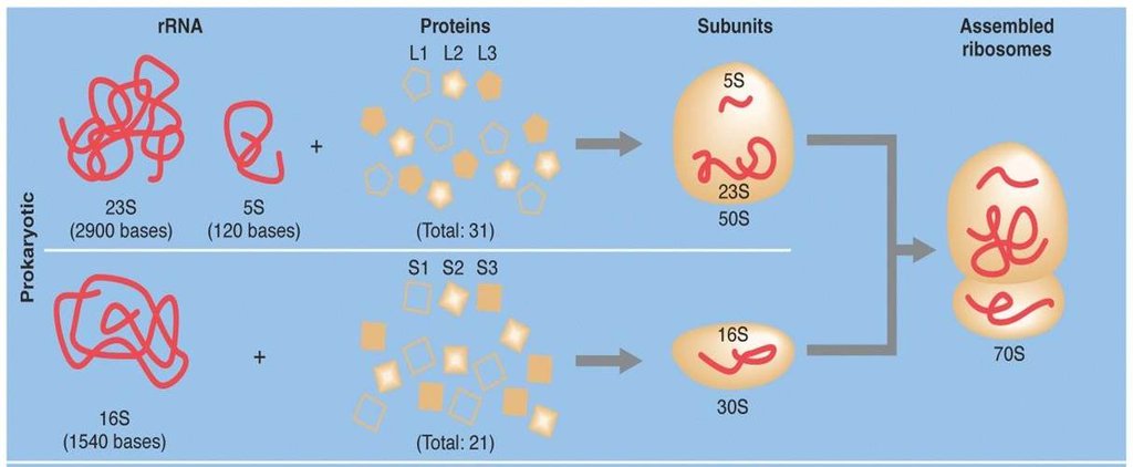
The Ribosomes are cell components that are present in all the cells i.e. both prokaryotes (e.g. bacteria) and eukaryotes (e.g. fungi, plants and animal)
- The ribosomes may be seen freely floating in cytosol or in groups attached to mRNA (polysome/polyribosome)
- They are not membrane-bound (no membrane surrounding them).
- They are made of rRNA (ribosomal RNA) and proteins.
- 70S ribosomes are present with 50S (larger) and 30S (smaller) subunits [S= Svedberg unit, which is a unit of sedimentation coefficient]
- The Smaller ribosomal subunit is made of 2 rRNA molecules (23S and 5S) and 31 proteins.
- The Larger ribosomal subunit is made of 1 rRNA (16S) and 21 proteins.
- These are the sites of protein synthesis (all the proteins in cell synthesized by them)
- Some of the ribosomes may be seen bound to the plasma membrane.
Examples of prokaryotic cells
Prokaryotic cells can be easily identified by the absence of the nucleus. All the bacteria, Mycoplasma, Cyanobacteria and PPLO (Pleuro Pneumonia Like Organisms) are included in prokaryotic organisms. Some of the examples of bacteria are
- Escherichia Coli
- Lactobacillus
- Bacillus subtilis
- Vibrio cholerae
- Salmonella Typhimurium
- Staphylococcus aureus
Bacteria as example of prokaryotic cell
- Prokaryotes are all single-celled organisms, most of which you know of as bacteria.
- For example, the famous (or infamous) Escherichia coli bacterium is a prokaryote, as is the streptococcus bacterium responsible for strep throat. The Streptomyces soil bacteria, from which the antibiotic streptomycin is derived, is also a prokaryotic organism.
Components of bacterial cell

A bacterial cell consists of following components:
- Cell envelope (glycocalyx, cell wall, plasma membrane)
- Cytoplasm (mesosome, ribosome, chromatophores)
- Nucleiod
- Plasmids
- Inclusion bodies (gas vacuoles, inorganic inclusions, food reserve)
- Flagella
- Pili
- Fimbriae
Types of chromosomes based on the position of centromere

Based on the position of centromere, chromosomes are of four types:
- Telocentric: Centromere terminal in the area of telomere.
- Acrocentric: Centromere inner to telomere.
- Submetacentric: Centromere submedian.
- Metacentric: Cetromere median.
Comments
Post a Comment