Difference between cerebrum and cerebellum
| Cerebrum | Cerebellum |
| It is the largest part of the brain, forming four fifths of its weight. | It is the second largest part of the brain, forming one eighths of its mass. |
| It is a portion of the forebrain. | It is a portion of the hindbrain. |
| It is the seat of the highest mental faculties which governs reasoning, learning, memory, intelligence. | It regulates and coordinates the contraction of skeletal muscles. |
| Cerebrum responds to heat, cold, pain, touch, light and pressure. | It maintains equilibrium and controls posture. |
| White matter does not form arbor vitae. | White matter form arbor vitae. |
Mammalian characters in human brain
- Olfactory lobes are small and solid.
- Cerebral hemispheres are quite large in size and divided into lobes.
- Corpus callosum is also found.
- Optic lobes are solid and further divided into corpora quadri-gemina.
- Pons varolii is present.
- Cerebellum is very much folded and solid.
Types of synapse
A synapse is a junction between the pre-synaptic neuron and a post-synaptic neuron, which may or may not be separated by a gap called the synaptic cleft.
There are two types of synapses on the basis of nature of the transfer of information.
Chemical synapses
There are two types of synapses on the basis of nature of the transfer of information.
Chemical synapses
- They transfer/relay the information through the chemicals
- These chemicals are neurotransmitter molecules.
- Each synaptic vesicle contains neurotransmitter chemical molecules such as Acetyl choline.
- The neurotransmitters thus released bind to their specific chemoreceptors present on the post-synaptic membrane of the dendron.
- This binding opens sodium ion channels that allow the entry of ions to generate a new potential in the post-synaptic neuron.
- Chemical synapses are slow and sturdy
- They can transmit signal only in one direction (post-synaptic to pre-synaptic )
- An electrical synapse is a junction in which the two neurons are connected by channel proteins
- They transfer/relay information through electrical signals
- They are faster as they do not receptors
- The signal can be transmitted in both the directions.
Reflex arc
- The path taken by the nerve impulses in a reflex is called a reflex arc.
1. Receptor- Protein molecule usually found embedded within the plasma membrane surface of a cell that receives chemical signals from outside the cell.
2. Sensory nerve- Carries the message from sensory neuron to spinal cord.
3. Relay centre- Intermediate neuron of spinal cord which transmits the impulse from sensory to motor neuron.
4. Motor nerve- Carries the message from spinal cord to effector organmuscle or gland.
5. Effector organ- Organ receives signal from motor nerve and act according to it.
Structure of reflex arc

The path taken by the nerve impulses in a reflex is called a reflex arc.
Components of a reflex arc:
1. Receptor- Protein molecule usually found embedded within the plasma membrane surface of a cell that receives chemical signals from outside the cell.
2. Sensory nerve- Carries the message from sensory neuron to spinal cord.
3. Relay centre- Intermediate neuron of spinal cord which transmits the impulse from sensory to motor neuron.
4. Motor nerve- Carries the message from spinal cord to effector organmuscle or gland.
5. Effector organ- Organ receives signal from motor nerve and act according to it.

Mechanism of vision
- The light rays from the object pass through the conjuctiva, cornea, aqueous humour, lens and vitreous humour in that order. All these structures refract the light such that it falls on the retina. This is called focussing.
- Maximum focussing is done by the cornea and the lens. The light then falls on the retina.
- This light is received by the photoreceptors - rods and cones, on the retina. The absorbed light activates the pigments present in the rods and cones.
- The pigments are present on the membranes of the vesicles. Thus, the light is then converted into action potentials in the membranes of the vesicles.
- These travel as nervous impulses through the rod or the cone cell and reach the synaptic knobs.
- From here the impulses are transmitted to the bipolar nerve cells, then to the ganglions and then to the optic nerves. Thus the nervous impulses generated in the retina are carried to the brain by about a million neurons of the optic nerve. The vision is controlled by the occipital lobe at the back of the brain. The information received is processed and we are able to see the image.
- The image formed on the retina is inverted. However, the brain makes us see the image erect. So, though the eyes are essential for vision, any damage to the optic nerves also results in impairment of vision.
Lens
- The lens is a transparent bifocal crystalline body located just behind the pupil.
- The lens is held in position by fibres called as the suspensory ligaments, which attaches it to ciliary body.
- The lens focusses the light from the outside world to form a tiny inverted image on the retina.

Eye and illusion
- An illusion is created when a stimulus is interpreted incorrectly. It is completely different from the external reality.
- The creation of illusion mainly depends on the way the receptor cells interacts with each other.
- Illusion can be generated by improper coordination between the optic lobe of the brain and receptors of an eye.
Sense organs for vision
- Eyes are the sense organs which is used for the sense of vision.
- It helps us to visualise the objects in the surroundings.
- In birds, it helps to estimate the distance and range of vision. Kites, eagles and vultures have a strong sense of vision.
- Eyes help us to differentiate amongst different colours.
Structure of ear

Structure of ear
- Ear has major three divisions.
- Outer ear consisting of pinna, auditory canal and ear drum.
- Middle ear consists of ear ossicles (malleus, incus, stapes), oval window, round window and inner opening of the Eustachian tube.
- Inner ear consists of semicircular canals, utriculus, sacculus and cochlea.
- External ear collects the sound waves.
- The hearing can be done according to the responses given by the brain.

Internal ear
Parts of internal ear are as follows:
- Cochlea: Contains a series of fluids, channels and membranes that transmit vibrations to the spiral organ, the organ of hearing, hair cell in the spiral organ produce receptor potentials, which elicit nerve impulses in the cochlear branch of the vestibulocochlear (VIII) nerve.
- Semicircular ducts: Conatin cristae, site of hair cell for dynamic equilibrium.
- Utricle and saccule: Contains macula, site of hair cells for dynamic equilibrium.
Organ of Corti
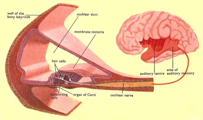
- The organ of Corti lies with the cochlea of the inner ear. In the organ of Corti, sound vibrations which pass along the cochlear duct are converted into nerve impulses.
- These impulses are transmitted along the cochlear nerve, or auditory nerve, to the brain, where they are interpreted as sound.
Mechanism of hearing
- The sound waves are directed towards the ear canal by the pinna. The waves that enter the canal are concentrated and made to strike against the tympanum.
From the basilar membrane the vibrations are picked up by the sensory hair cells of the organ of corti and transmitted as action potentials to the neurons of the auditory nerve fibres. The exact mechanism of transformation of the sound waves into the action potentials is not known. The action potentials are then transmitted as nerve impulses to the auditory cortex of the brain through the auditory nerve. - The vibrations are picked up by the malleus on the other side. These vibrations are transmitted to the fenestra ovalis via the incus and the stapes.
- The vibrations that strike the oval window are amplified 22 times more than those that struck the tympanum.
- These vibrations travel along the vestibular canal to the end of the cochlea and then to the tympanic canal.
- The vibrations are also transmitted via the Reisnner's membrane to the basilar membrane and then to the tympanic canal.
- Note that the vibrations travel along the vestibular and tympanic canals in the opposite directions.
Maintenance of equilibrium
- Membranous labyrinth are the structure of equilibrium.
- Whenever the animal gets tilted the hair cells of the cristae and maculae are stimulated by the movement of the endolymph and otolith.
- The impulse is carried to the brain and the change of position is detected by the medulla oblongata of the brain.
- After which, the brain sends impulses to the muscles to regain the normal conditions.
Mechanism of hearing
- Sound waves sets the eardrum into vibration.
- Vibrating eardrum sets ear ossicles malleus (hammer), incus (anvil) and stapes (stirrup) into motion.
- Vibrating stirrup transmits vibrations to oval window, then to cochlear canal, stimulates the sense cells of a cochlea, impulses through the auditory nerve and reach the brain.
Comments
Post a Comment