Tissue
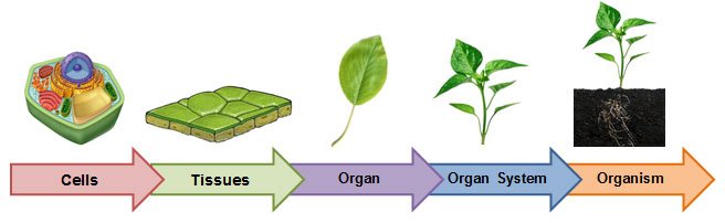
- Tissue is a group of cells that are similar in structure and are organised together to perform vital functions.
- Different types of tissues work together and form an organ.
- Organs are arranged into a system to form an organ system. For example, reproductive system etc.
- An organism consists of such organ system working together.
- Study of tissues is called as histology.
Simple permanent tissue (supportive tissue) - Sclerenchyma

- These are long, thick, narrow, dead cells with a deposit of lignin in their cell wall.
- They have no intercellular spaces.
- Sclerenchyma occur around the vascular tissues in stems, in the veins of leaves, and in the covering of seeds and nuts.
- They provide strength to the plant.
Types of xylem
The different components of the xylem include tracheids, vessels, xylem parenchyma and xylem fibres.
- Tracheids: Provide support to the plant.
- Xylem vessels: Allows transport of water and minerals vertically.
- Xylem parenchyma: Stores food and helps in sideways conduction of water and minerals.
- Xylem fibres: Supports the plant.
Types of phloem

The different elements of phloem include sieve tubes, companion cells, and phloem parenchyma and phloem fibres.
- Sieve tubes: Transport sugars and nutrients up and down the plants in sieve cells.
- Companion cells: Controls the activity of sieve tube.
- Phloem parenchyma: Provides mechanical strength to the plant.
- Phloem fibres: Stores compound such as starch.
Ground tissue system
- Ground tissue system forms the main bulk of the plant body and it extends from below the epidermis to the centre.
- It can be distinguished into cortex, pericycle, pith and medullary rays.
- In monocotyledonous stems, where vascular bundles are scattered, there is no distinction in the ground tissue system.
Types of vascular bundles
Xylem is classified on the basis of the position of protoxylem:
- Endarch is the arrangement where protoxylem (older xylem) is inside and metaxylem (newly formed) is outside.
- Exarch xylem has metaxylem towards the centre and protoxylem is present towards the periphery.
- Mesarch has developing xylem towards both sides which leads to the formation of protoxylem in the middle and protoxylem on the both sides.
- Conjoint- In this the xylem and phloem are present on the same radius. It is majorly of two types: Collateral vascular bundle is that in which the phloem and xylem lie on the same radius, with the phloem located toward the periphery of the stem and the xylem toward the centre. In bicollateral, two cambium and two phloem are present on both sides of xylem.
- Concentric vascular tissues have one of the tissue as central core surrounded by the other.
Anatomy of dicot root

The transverse section of the dicot root shows the following plan of arrangement of tissues from the periphery to the centre.
A. Pericycle
- Rhizodermis or epiblema:
- The outermost layer is made up of single layer of parenchymatous cells without intercellular spaces. Stomata and cuticle are absent.
- Root hairs are always single celled.
- Cortex consists of oval or rounded loosely arranged parenchymatous cells.
- These cells may store food reserves.
- It is made up of single layer of barrel shaped parenchymatous cells.
- The radial and the inner tangential walls of endodermal cells are thickened with suberin. These thickenings are known as casparian strips.
- But these casparian strips are absent in the endodermal cells which are located opposite to the protoxylem elements.
A. Pericycle
- Pericycle is generally a single layer of parenchymatous cells found inner to the endodermis. Lateral roots originate from the pericycle.
- Vascular tissues are in radial arrangement.
- The tissue by which xylem and phloem are separated is called conjunctive tissue.
- Xylem showes exarch and tetrarch condition .
- Metaxylem vessels are generally polygonal in shape
Anatomy of dicot stem

1. Epidermis
- It is a protective outermost single layer of parenchymatous cells without intercellular spaces.
- The outer walls of the epidermal cells have a layer called cuticle and multicellular hairs (trichomes).
- Below the epidermis, cortex is differentiated into few layers of collenchyma cells that make hypodermis which gives mechanical strength to the stem.
- A few layers of chlorenchyma cells are present with conspicuous intercellular spaces. Some resin ducts also occur here.
- The third zone is made up of parenchyma cells. These cells store food materials.
- The cells of this layer are barrel shaped arranged compactly without intercellular spaces.
- Due to abundant starch grains in these cells, this layer is also known as starch sheath.
- It consists of pericycle, vascular bundles and pith.
- Pericycle occurs between the endodermis and vascular bundles in the form of a few layers of sclerenchyma cells.
- In dicot stem, vascular bundles are arranged in a ring around the pith.
- Each vascular bundle is conjoint, collateral, open and endarch.
- The large central portion called pith composed of parenchyma cells with intercellular spaces.
- The extension of pith between vascular bundles are called as pith ray or medullary rays.
- Function of the pith is storage of food.
Dorsiventral mesophytic leaf
A transverse section through the midrib region of a typical dorsi-ventral leaf (sunflower) reveals the following structure.
- Epidermis is in two layers, one on each surface of the leaf. Both the layers are composed of compactly arranged, barrel-shaped cells. Intercellular spaces are absent. A cuticle surrounds both the layers. Multicellular hairs called trichomes are present on both the layers. Stomata occur only in the lower epidermis. This condition is described as hypostomatic.
- Mesophyll is the ground tissue that occurs between the two epidermal layers. It is exclusively composed of chlorenchyma cells. The mesophyll is characteristically differentiated into two regions namely, an upper palisade parenchyma and a lower spongy parenchyma.
- Veins represent the vascular bundles. They are found irregularly scattered in the mesophyll due to reticulate venation. The largest and the oldest vein is found in the centre. It is known as midrib vein.
Anatomy of primary monocot root

1. Rhizodermis or epiblema
A. Pericycle:
- The outermost layer of parenchymatous cells without intercellular spaces. Stomata and cuticle are absent.
- Root hairs are always single celled.
- The cortex is homogenous, consists of oval or rounded loosely arranged parenchymatous cells.
- The function of cortical cells is storage.
- It is made up of single layer of barrel shaped parenchymatous cells.
- The radial and the inner tangential walls of endodermal cells are thickened with suberin. These thickenings are known as casparian strips.
- But these casparian strips are absent in the endodermal cells which are located opposite to the protoxylem elements.
A. Pericycle:
- A single layer of parenchymatous cells found inner to the endodermis. Lateral roots originate from the pericycle.
- Vascular tissues are in radial arrangement.
- Xylem and phloem are separated by sclerenchymatous conjunctive tissue.
- Xylem shows exarch and polyarch condition.
- Metaxylem vessels are generally circular in shape.
- The central portion is occupied by a large pith consist of thin walled parenchyma cells with intercellular spaces.
- These cells are filled with abundant starch grain.
Anatomy of monocot stem

1. Epidermis
- It is the outermost layer made up of single layer of tightly packed parenchymatous cells with thick cuticle.
- There are no epidermal outgrowths.
- A few layer of sclerenchymatous cells lying below the epidermis constitute the hypodermis, gives mechanical strength to the plant.
- It is not differentiated into cortex, endodermis, pericycle and pith.
- The ground tissue is represented by several layers of loosely arranged parenchyma cells enclosing prominent intercellular spaces.
- The ground tissue is meant for storage of food.
- Vascular bundles are scattered in the parenchymatous ground tissue.
- Vascular bundles are numerous, small and closely arranged in the peripheral portion.
- Towards the centre, the bundles are comparatively large in size and loosely arranged.
- Each vascular bundle is surrounded by a sheath of sclerenchymatous fibres called bundle sheath.
- The vascular bundles are conjoint, collateral, endarch and closed.
- The phloem in the monocot stem consists of sieve tubes and companion cells.
- Phloem parenchyma and phloem fibres are absent.
- The two metaxylem vessels are located at the upper two arms and one or two protoxylem vessels at the base. (Y shaped)
- In a mature bundle, the lowest protoxylem disintegrates and forms a cavity known as protoxylem lacuna.
Isobilateral leaf

A transverse section passing through the midrib region of an iso-bilateral leaf (maize) reveals the following structure.
- Epidermis is in two layers, one on each surface of the leaf. Both the epidermal layers are composed of compactly arranged, barrel shaped cells. Cuticle and trichomes are present in both the layers. Stomata are found in both the epidermal layers. This condition is described as amphistomatic. A few cells in the upper epidermis are enlarged to form motor cells called bulliform cells.
- Mesophyll is ground tissue that occurs between the two epidermal layers. It is composed of many layers of loosely arranged, spherical or oval chlorenchyma cells. Intercellular spaces are prominent.
- Veins are found parallely arranged in the mesophyll (parallel venation). Each vascular bundle is surrounded by a bundle sheath composed of a single layer of compactly arranged barrel-shaped cells. The bundle sheath encloses both phloem and xylem. Phloem is found towards lower epidermis and xylem towards upper epidermis. In the xylem, only two protoxylem and two metaxylem vessels are present. The vascular bundle is described as conjoint and collateral with endarch xylem.

Secondary growth in dicot root
- The vascular cambium is entirely secondary in origin.
- The cell is located just below the phloem bundles and cells of pericycle above the protoxylem region, regain the capacity to divide and a wavy ring is formed which later becomes circular.
- Secondary xylem is produced inside and secondary phloem outside like stem and the diameter of root increases.
Comments
Post a Comment