Process of digestion
- Food has several components like carbohydrates, proteins, vitamins, minerals and fats.
- The components of food are digested in the stomach with the help of digestive juices.
- The digestive juices act on the food in the stomach and break into simpler compounds.
- The digestive juices are acidic.
Tongue

Tongue is a muscular organ. It is freely movable at the front end. The tongue is attached to the floor of the mouth by a fold called the frenulum linguae. An inverted v-shaped furrow, termed the sulcus terminalis is present across the tongue.
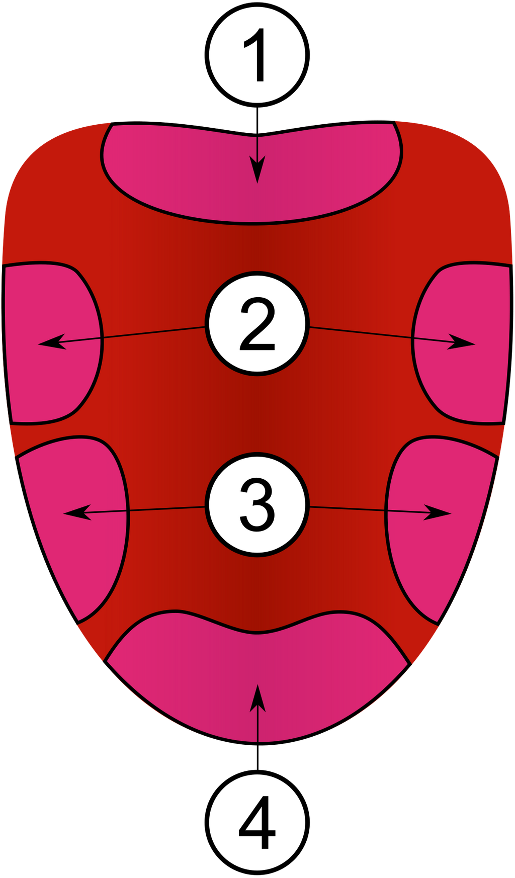
The tongue has many papillae on its surface. These papillae contain taste buds and are of 4 types:
The tongue has many papillae on its surface. These papillae contain taste buds and are of 4 types:
- Vallate papillae- They are large in size and are 8 to 10 in number. They lie near the base of the tongue. Each of them are surrounded by circular grooves. The walls of these groove contain taste buds.
- Filiform papillae- They are the smallest in size amongst all the papillae.
- Fungiform papillae- They are round and red in color. The epithelium of this papillae have taste buds.
- Foliate papillae- The tongue has 4 to 5 transverse folds at the border and near the base. They have many projections.
- Helps to taste food. The taste buds are sensitive to salty, sour, bitter and sweet tastes.
- It helps in chewing and pushing the bolus backward.
Human teeth
Dental formula is used to calculate the number of teeth in mammals. The human dental formula is as follows:
- Human child (upto 2 years):
Thus, the total number of teeth are = 2x10= 20 (milk teeth) - Human adolescent (17-20 years):
Thus, the total number of teeth are = 2x14= 28 (permanent teeth) - Human adult:
Thus, the total number of teeth are = 2x16= 32 (permanent teeth + wisdom teeth)
Dental formula
Dental formulae indicate the number of each type of tooth in a given species.
The jaw is bilaterally symmetrical. Hence, the dental formulae represent the number considering the half jaw only.
The numerator of the fraction represents the number of teeth in the upper jaw and denominator represents the number of teeth in the lower jaw.
The formula represents number of teeth in the following order (left to right): Incisors, Canine, Premolars, Molars
Example: =
Difference between different types of teeth
| Type of tooth | Location | Feature | Function | |
| 1. | Incisor | Incisors are situated in the centre of both, top and bottom jaw. |
| They are adapted for cutting. |
| 2. | Canine | Located beside the incisors. |
| They are used for tearing food. |
| 3. | Premolar | They are located beside canines. |
| Premolars are used to chew and tear food. Initial grinding is done by them. |
| 4. | Molar | They are located at the back of the mouth. |
| Molars are responsible for crushing and grinding the food. |
Milk teeth and Permanent teeth
Teeth are present in the buccal cavity and help to churn food.
The first set of teeth grows during infancy and they fall off at the age of 6-8 years are known as milk teeth.
The second set of teeth which remains for rest of the life are known as permanent teeth.
The different types of permanent teeth are molar, premolar, canine and incisor.
The first set of teeth grows during infancy and they fall off at the age of 6-8 years are known as milk teeth.
The second set of teeth which remains for rest of the life are known as permanent teeth.
The different types of permanent teeth are molar, premolar, canine and incisor.
Parts of a tooth

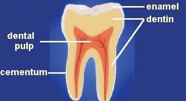
Each tooth consists of four parts:
- Enamel: It is the hardest substance in the body. Enamel is the hard, white outer covering of the tooth. It is mostly made of calcium phosphate, a rock-hard mineral.
- Dentine: A layer underlying the enamel. It is made of a hard calcified tissue. Dentine encloses the pulp cavity.
- Pulp: Pulp is a part of pulp cavity. It is a soft and gelatinous connective tissue. Blood vessels and nerves run through the pulp of the teeth.
- Cementum: A layer of connective tissue that binds the roots of the teeth firmly to the gums and jawbone.
Incisors

Incisors are situated in the center of both- top and bottom jaw. They have the following features:
- There are 4 incisors in each jaw.
- They are thin, flat-bottom, narrow- edged and sharp in shape.
- Incisors are mainly adapted for cutting.
- Incisors help us to make the initial bite.
Types of teeth

The teeth are classified into different types on the basis of its use. They are mainly used for cutting, biting, tearing, chewing and grinding.
They are differently shaped permanent teeth to break the food into small chunks which make the digestion easy.
Permanent teeth are divided into four types. Each has special structure and functions. They are:
They are differently shaped permanent teeth to break the food into small chunks which make the digestion easy.
Permanent teeth are divided into four types. Each has special structure and functions. They are:
- Incisors
- Canine
- Premolars
- Molars
Dental formula in humans
Dental formula is used to calculate the number of teeth in mammals. The human dental formula is as follows:
- Human child (upto 2 years):
Thus, the total number of teeth are = 2x10= 20 (milk teeth) - Human adolescent (17-20 years):
Thus, the total number of teeth are = 2x14= 28 (permanent teeth) - Human adult:
Thus, the total number of teeth are = 2x16= 32 (permanent teeth + wisdom teeth)
Dental formula
Dental formulae indicate the number of each type of tooth in a given species.
The jaw is bilaterally symmetrical. Hence, the dental formulae represent the number considering the half jaw only.
The numerator of the fraction represents the number of teeth in the upper jaw and denominator represents the number of teeth in the lower jaw.
The formula represents number of teeth in the following order (left to right): Incisors, Canine, Premolars, Molars
Example: =
The jaw is bilaterally symmetrical. Hence, the dental formulae represent the number considering the half jaw only.
The numerator of the fraction represents the number of teeth in the upper jaw and denominator represents the number of teeth in the lower jaw.
The formula represents number of teeth in the following order (left to right): Incisors, Canine, Premolars, Molars
Example: =
Salivary glands
Saliva is a watery substance located in the mouths of humans and animals, secreted by three pairs of salivary glands, parotid, submandibular and sublingual gland.
Functions of saliva:
Functions of saliva:
- It helps in lubricating the inner lining of the mouth making it easier for swallowing and speaking.
- It acts as a solvent.
- It helps in forming bolus.
- Allows digestion of starch.
Tongue

The tongue is a muscular organ.
It is attached to the back of the buccal cavity. In the front, it can move freely in all directions.
The function of the tongue is to push the food forward and help us to swallow it.
It is also used to taste food.
Food can be tasted with the help of taste buds present on the tongue.
It is attached to the back of the buccal cavity. In the front, it can move freely in all directions.
The function of the tongue is to push the food forward and help us to swallow it.
It is also used to taste food.
Food can be tasted with the help of taste buds present on the tongue.
Mouth (Buccal cavity)
- The buccal cavity is the first cavity of the alimentary canal which includes teeth, tongue, and palate (Palate is the roof of the mouth. It separates the mouth and the nose.).
- The buccal cavity helps to take the food inside by the process of ingestion (ingestion is the process of taking food into the body).
Oesophagus

The esophagus or the food pipe is a muscular tube and has the following features:
- It starts from the pharynx to the stomach. The pharynx is present behind the trachea.
- Food/bolus is pushed through the esophagus into the stomach by means of a series of contractions called peristalsis. These contractions occur in the muscles in its wall.
- There is no digestion in this part.
- It is also called as gullet.
- The opening of the esophagus into the stomach is regulated by a muscular sphincter called gastro-esophageal sphincter.
Stomach

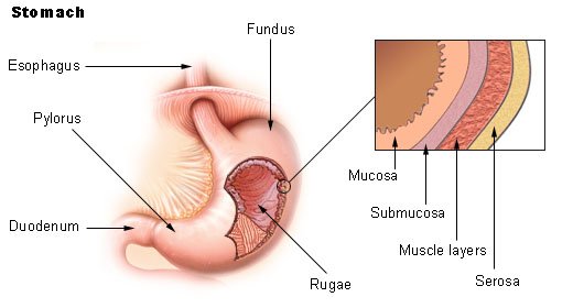
Structure of the stomachIt is a highly muscular, J-shaped organ.
It is located in the upper left portion of the abdominal cavity.
The stomach has three parts:
The innermost layer lining the lumen is the mucosa. This layer forms irregular folds called rugae in the stomach.
Mucosa form gastric glands in the stomach.
Function of the stomach
The gastric glands present in its walls secrete gastric juice containing hydrochloric acid (HCl) and enzymes, like pepsinogen.
HCl activates pepsinogen into pepsin and kills bacteria.
Proteins are broken into smaller fragments called peptones by the enzyme called pepsin.
It eventually leads to the formation of chyme, which is moved to the intestine through pylorus.
It is located in the upper left portion of the abdominal cavity.
The stomach has three parts:
- Cardiac portion: The esophagus opens here.
- Fundic region
- Pyloric portion: It opens in the first part of small intestine.
The innermost layer lining the lumen is the mucosa. This layer forms irregular folds called rugae in the stomach.
Mucosa form gastric glands in the stomach.
Function of the stomach
The gastric glands present in its walls secrete gastric juice containing hydrochloric acid (HCl) and enzymes, like pepsinogen.
HCl activates pepsinogen into pepsin and kills bacteria.
Proteins are broken into smaller fragments called peptones by the enzyme called pepsin.
It eventually leads to the formation of chyme, which is moved to the intestine through pylorus.
Blood supply to the liver
The liver receives blood through two routes; the hepatic artery and the hepatic- portal vein.
Hepatic artery
Hepatic artery
- supplies oxygenated blood to the liver.
- comes from the celiac trunk of the abdominal aorta and bifurcates (divides) into two branches; right hepatic artery the right and the left hepatic artery
- Right hepatic artery enters the right half of the liver and the left hepatic artery enters the left half.
- The right hepatic artery further divided into cystic artery (supplies oxygenated blood to gallbladder and cystic duct)
- Supplies deoxygenated blood to the liver
- 75% of blood reaching the liver is supplied by the hepatic portal vein.
- The blood in the hepatic vein is rich in nutrients and toxins as it is connected to the capillaries of the gastrointestinal tract.
- The hepatic vein in the liver is divided into many capillaries called sinusoids.
- In the sinusoids, nutrients are absorbed and toxins are neutralized (converted into less harmful chemicals).
Protein digestion in small intestine
- Pancreatic juice contains proenzymes trypsinogen, chymotrypsinogen and procarboxypeptidase and enzyme elastase.
- The bile provides alkaline medium for various reactions.
- The intestinal juice contains aminopeptidases and dipeptidases, and enterokinase or enteropeptidase.
- Out of these enterokinase activates the trypsinogen. Aminopeptidase hydrolyses the terminal amino group from peptide bonds to release the last amino acid from the peptides thus making the peptide shorter.
- Dipeptidase acts on dipeptides to release the individual amino acids.
Lacteal

A lacteal is a lymphatic vessel. It is present in a villus of the small intestine. Its function is to move lymph through the intestines. This can help to keep lymph circulating through the small intestine. They also help in transferring nutrients from the small intestine into the blood. Fats in the form of chylomicrons are transported into the lacteals by exocytosis.
Histology of human alimentary canal

The wall of the alimentary canal (from the esophagus to rectum) consists four basic layers:
- Serosa: It is the outer mode layer. Serosa is made up of thin mesothelium (epithelium of visceral organs) with areolar connective tissue.
- Muscularis: It is composed of smooth muscles usually arranged into an outer longitudinal and inner circular muscle fibre.
- Submucosa: It is composed of loose connective tissue richly supplied with blood and lymphatic vessels and in some areas with glands.
- Mucosa: It is the inner most layer which secretes mucus. It is composed of three layers:
- Muscularis mucosa- It is a thin layer of smooth muscles.
- Lamina propria- It is a layer of connective tissue. It is unusually cellular so it is compared to most connective tissues.
- Epithelium- innermost layer and is responsible for most digestive, absorptive, and secretory processes.
Good food for digestion
- Carbohydrates, fats, proteins, vitamins and minerals are the major components of the food.
- These components are required in the desired amount which helps in the proper growth and development.
- Good food provides a high percentage of nutrients which helps to strengthen our body and helps to protect us from several diseases.


Comments
Post a Comment